


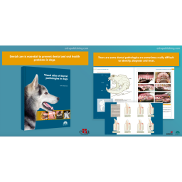
Dental care is essential to prevent dental and oral health problems in dogs. It is nowadays a routine procedure for most veterinary practices. However, some dental pathologies are sometimes really difficult to diagnose and treat. With almost 400 high-quality images, clear anatomical illustrations, and descriptions of numerous diagnostic tests, this atlas constitutes a reference handbook for a quick visual identification with a clear orientation towards a precise diagnosis and treatment. In short, this is an essential tool for a better understanding of dental pathologies in dogs.
"This Atlas constitutes a new and valuable contribution to the literature of Veterinary Dentistry. It is not a textbook; rather it uses a wide spectrum of well-organized visual material to clearly explain different pathologies in dogs.
The Spanish author has selected well-known international collaborators to contribute to a comprehensive range of high-quality visual material comprising almost 400 pictures arranged on more than 100 pages. The chapters and pages are laid out in a clear and interesting format that communicate information effectively and retain the reader's interest on a voyage of discovery.
The overall organization in terms of etiology, clinical, and diagnostic features together with well-labeled diagrams and illustrations is a format that allows the reader to quickly delve down to whatever level of detail is required. In particular, the juxtaposition of different visual formats provides a multidimensional insight. For example, well-selected clinical photographs and matching radiographs highlight the value of radiography in diagnosis.
The Atlas is both a well-conceived idea and a well-executed project. This one book has such broad coverage of dental pathologies in dogs that it constitutes a uniquely valuable addition to the library of any veterinary surgeon."
Authors:
JAVIER COLLADOS SOTO
Javier Collados Soto graduated in Veterinary Medicine from the Complutense University of Madrid (UCM) in 1994. Specialising exclusively in Veterinary Dentistry and Oral Surgery, he works in numerous veterinary practices and hospitals in Spain, concentrating his services in Madrid. He is responsible for the Dentistry and Oral Surgery service at the Sierra de Madrid Veterinary Hospital. He was a lecturer and subject coordinator in Animal Dentistry at the Faculty of Veterinary Medicine of the Alfonso X el Sabio University of Madrid. Always showing his interest for his specialisation, he has had several stays at the Dentistry and Oral Surgery Service of the University of California (UCDavis) Veterinary Medical Teaching Hospital, USA. He has been member of the European Veterinary Dental Society (EVDS) since 1999 and he is also one of the founder members of the Spanish Society of Veterinary Experimental Dentistry and Maxillofacial Surgery (SEOVE, Sociedad Española de Odontología-Cirugía Maxilofacial Veterinaria y Experimental). He has published many articles in this specialty and has participated as a speaker in conferences and national and international courses in the field of Veterinary Dentistry.
Table of Contents:
1. introduction
dental positioning terms
histological structure of the tooth in dogs
and cats
dog and cat teeth diagrams
example of periodontal probing
Classification of periodontal diseases
(AVdC, 2007)
Classification of the bacterial plaque
and dental calculus indices
(Logan & boyce, 1994)
Classification of the gingival index
(Wolf et al., 2005)
Classification of tooth mobility
(AVdC, 2007)
Classification of furcation defects
(AVdC, 2007)
Classification of dental fractures
(AVdC, 2007)
endoperiodontal lesions
Classification of dental resorptions
(AVdC, 2007)
2. dental and oral cavity
pathologies in the dog
oral pathology in dogs
permanent teeth
permanent teeth
deciduous teeth
Normal tooth eruption
dog dental radiography models
dental abrasion
Alteration of dental development and eruption
dental agenesis
unspecific tooth alterations
Non-physiological diastema
impacted teeth
impacted (embedded) teeth
incomplete eruption
Fusion
germination
hyperdontia (supernumerary teeth)
hypodontia
hypoplasia-hypomineralization of enamel
microdontia
odontodysplasia
oligodontia
dentigerous cyst
iatrogenic alterations
pathological attrition
dental absence
dental caries
dental discoloration
dental fracture
enamel fracture
uncomplicated crown fracture
Complicated crown fracture
Complicated crown-root fracture
Complicated crown-root fracture
root fracture
dental luxation-subluxation
pulp pathology
dental resorption
dental stains
3. references
4. Alphabetical index
Fiche technique
Références spécifiques
This atlas shows Cardiology in a way it had never been done before. The images it contains, of both the healthy heart and the main heart diseases that affect dogs and cats, have a unique technical and artistic value.
Electrocardiography of the dog and cat, 2nd edition provides all the theoretical and practical information necessary to understand and identify rhythm abnormalities. This text will be particularly useful to accurately diagnose complex arrhythmias using precise electrocardiographic criteria that the authors have developed by confirming mechanisms of arrhythmias with intracardiac electrophysiologic studies. This book is a must-have reference for anyone interested in learning the basics of electrocardiography and those who face the challenge of interpreting the most complex arrhythmias in a clinical or research setting.
This new volume of the Pet owner educational atlas collection on the cardiorespiratory system of dogs and cats pursues the same objective as the previous books: helping veterinary surgeons to communicate with pet owners. In addition, as its contents are focused on one single system, the specificity of the topics addressed will also help veterinary practitioners to consolidate and broaden their knowledge.
This new volume of the collection “Small animal surgery” is a selection of the main surgical procedures explained in the previous volumes. The greatest asset of this book is that it is based on high-quality videos, which accompany each of the surgical techniques. These are classified according to the degree of difficulty. This book is the result of the author’s thorough and careful work and is an essential resource both for veterinary professionals in practice and students of veterinary surgery.
Photographs, diagrams and real clinical cases explain, step by step, the methods of approach and resolution for the main surgical interventions in the rectal, anal and perineal zones. An essential work for both practicing professionals and surgery students.
Dental care is essential to prevent dental and oral health problems in dogs. It is nowadays a routine procedure for most veterinary practices. However, some dental pathologies are sometimes really difficult to diagnose and treat. With almost 400 high-quality images, clear anatomical illustrations, and descriptions of numerous diagnostic tests, this atlas constitutes a reference handbook for a quick visual identification with a clear orientation towards a precise diagnosis and treatment. In short, this is an essential tool for a better understanding of dental pathologies in dogs.


